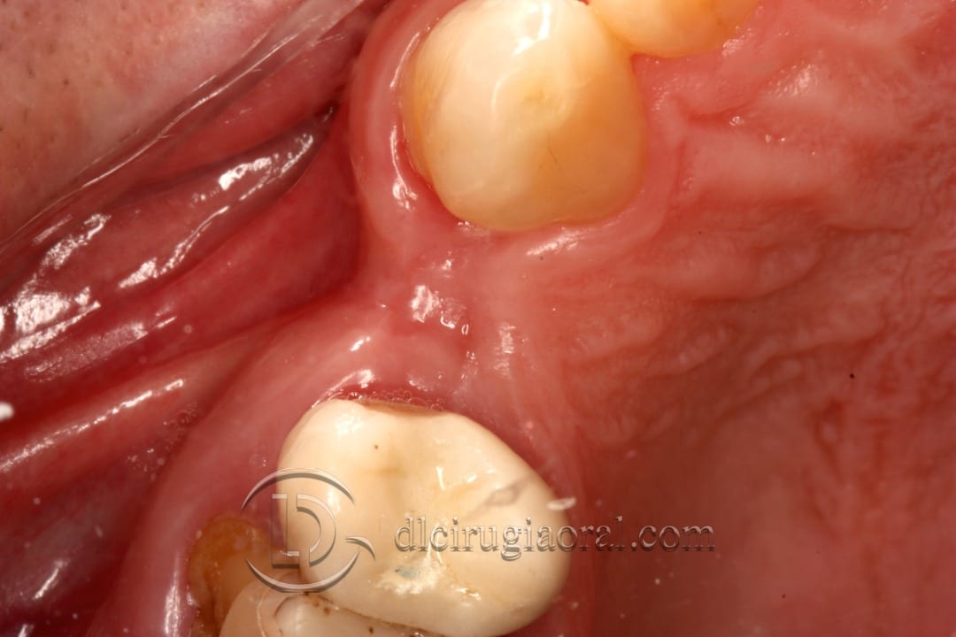
We present the case of a 55-year-old patient, medically healthy, referred for assessment and treatment of a horizontal defect in the upper molar area. The main concern involved the upper right premolar region, where the patient needed a dental implant.
Clinical and radiographic examination revealed a horizontal bone defect, caused by a traumatic extraction that had led to loss of the buccal plate. This defect compromised the ridge volume and required bone augmentation before or during implant placement.
Reconstructing the horizontal defect in the upper molar region
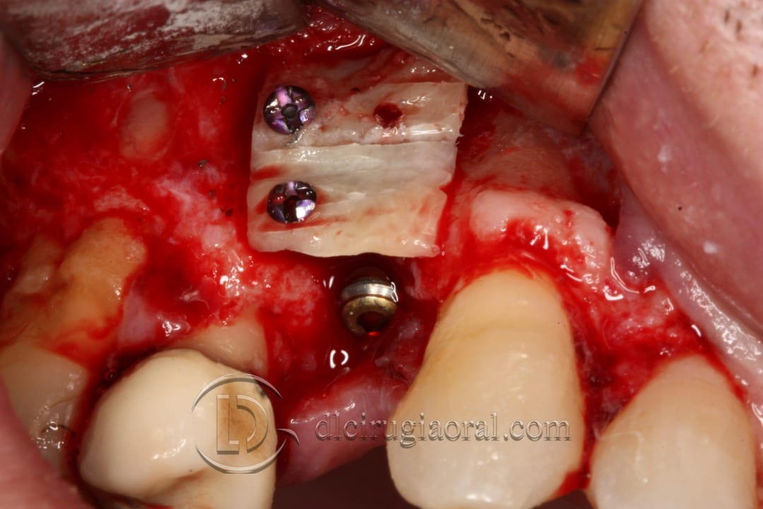
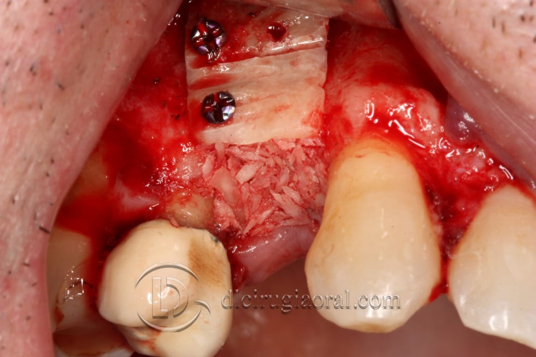
To restore the defect, we opted for a crestal approach. This allowed us to place the implant in the ideal prosthetic position while simultaneously grafting the defect.
We harvested a cortical block from the mandibular external oblique ridge, dividing it into two thin plates for better vascularization. The internal space of the graft was filled with autogenous particulate bone, enhancing integration and volume stability.
Second surgery and additional implant placement
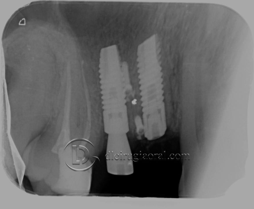
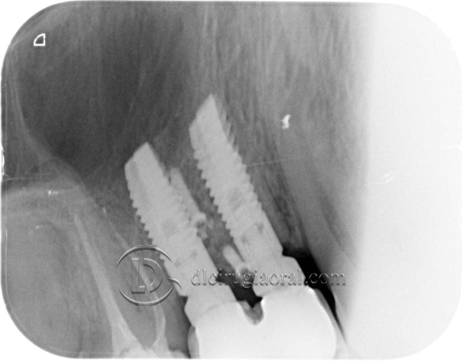
One month later, the patient returned due to a vertical fracture in the adjacent tooth (#15). We placed a second implant in that position. Unfortunately, no images were taken during that procedure.
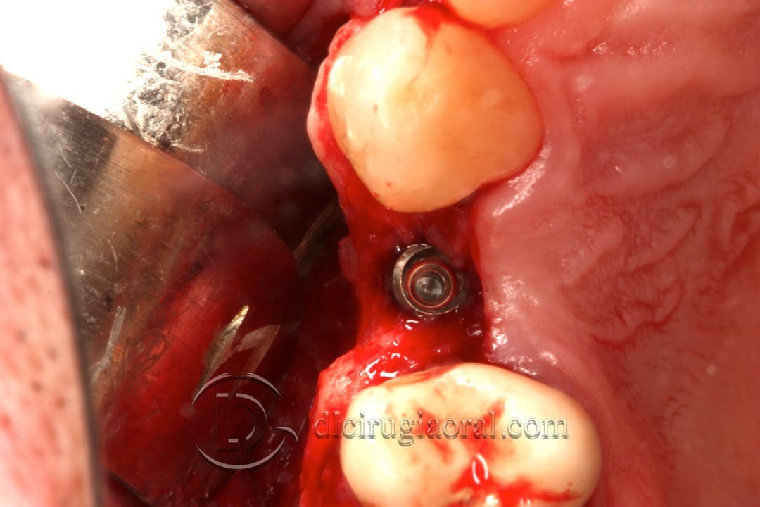
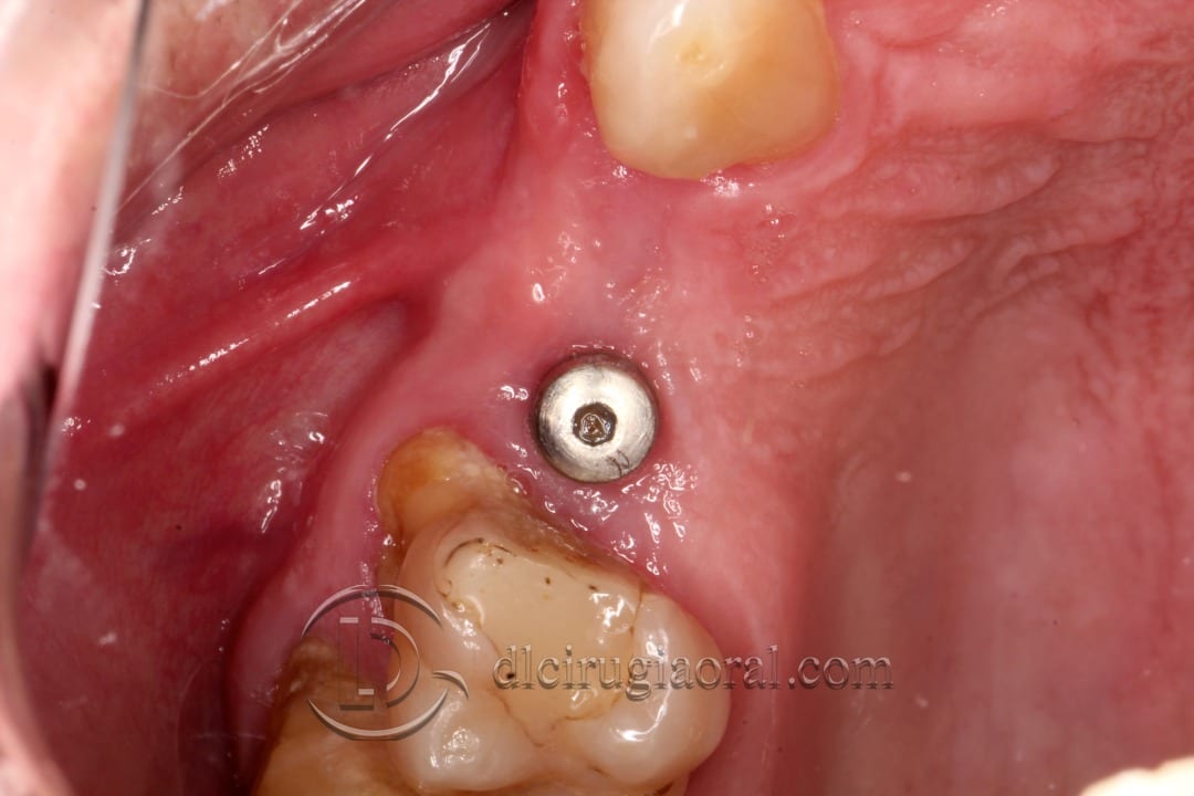
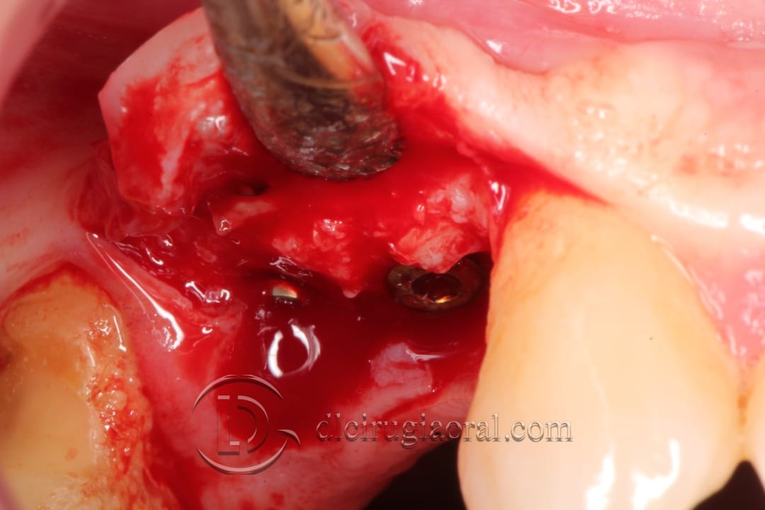
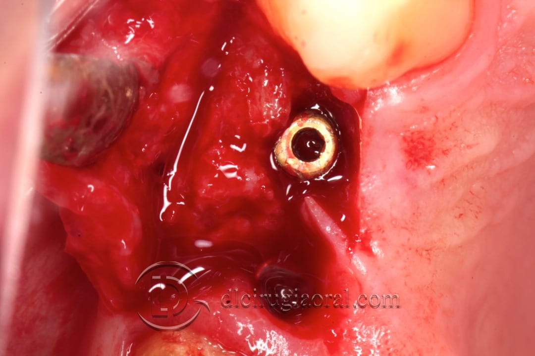
After 3 months, we performed the second-stage surgery. The regenerated site showed excellent bone volume and density. The horizontal defect appeared fully resolved, with stable integration of the implant.
Final prosthesis and follow-up
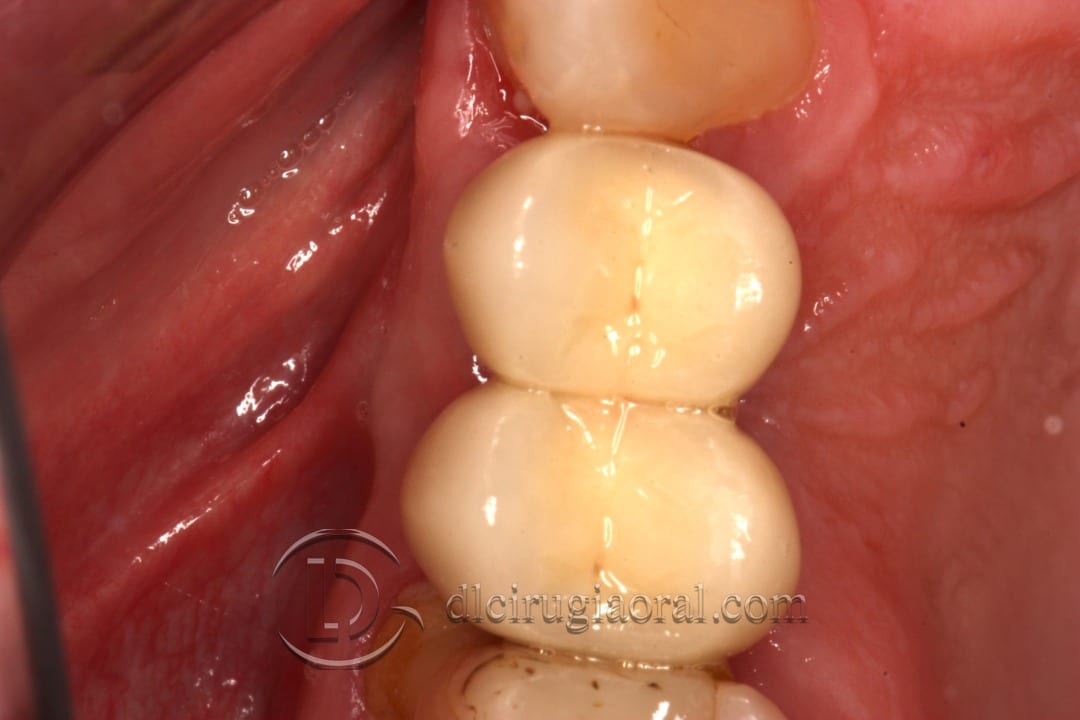
The prosthodontist at the referring clinic completed the case with a functional and esthetic prosthesis.
📎 To see more cases like this horizontal defect in upper molar, visit our clinical archive for detailed surgical documentation and outcomes.
Below, you’ll find photos of the original surgery, second-stage exposure, and final restoration.