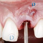

Drilling implant bed
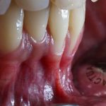
As we saw in the previous post, which you can view here, we reconstructed a posterior mandibular defect with a height of 2.4 millimetres to the dental nerve.
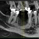
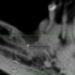
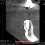
After the mandibular reconstruction, the patient became pregnant, and came back to us after one year. We requested a cone beam CT prior to implant placement, where we observed that the height behind the graft was 11.8 millimetres, so we decided to place 10mm long implants after gaining 9 millimetres with our reconstruction.
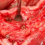
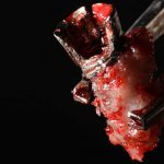
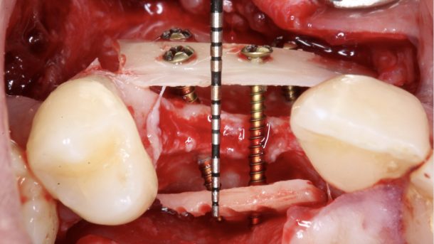
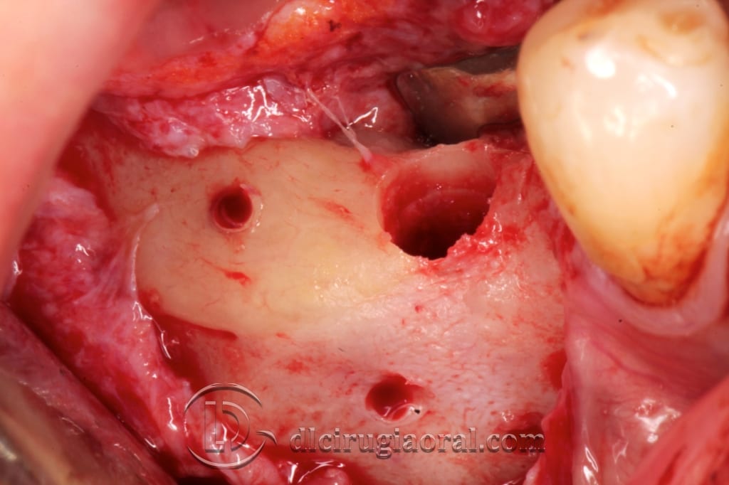
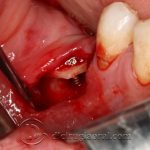
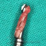
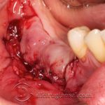
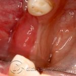
As you can see, we used a Kazanjian vestibuloplasty access to gain adhered gum. We got to the graft, where we observed its fixing screws with barely any reabsorption. We measured this reabsorption to see if there were exposed coils in these screws and drilled the grafted bone. This had a normal consistency and was vascularised.
We placed two 10 millimetre high implants and sutured the vestibuloplasty. You can observe from the tomographic cuts the previous situation, one year after grafting, and after placing implants.
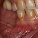
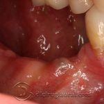
Finally, you can see that thanks to the vestibuloplasty we obtained adhered gum and bone plate to protect the implants.
-

- Previous image
-
- Reconstruction in tunnel
-
- After one year
-
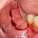
- Vestibuloplasty access
-

- Image of the graft
-

- Drilling the implant bed
-
- Appearance of grafted bone
-
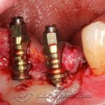
- Placement of implants
-
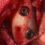
- Occlusal view
-
- Vestibuloplasty sutured
-
- Previous CT
-
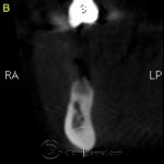
- Previous CT
-
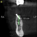
- Post-reconstruction CT
-
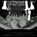
- Previous panoramic
-
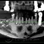
- Post-reconstruction panoramic
-
- Mesial implant cut
-
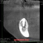
- Distal implant cut
-
- Healed vestibuloplasty occlusal view
-
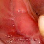
- Healed vestibuloplasty
Click to rate this post!
[Total: 0 Average: 0]