
Occlusal view
After the reconstruction seen in this post: Reconstruction following failed implant and graft, we waited 4 months and proceeded to open said graft for the implant surgery.
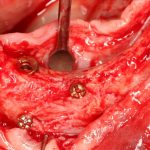
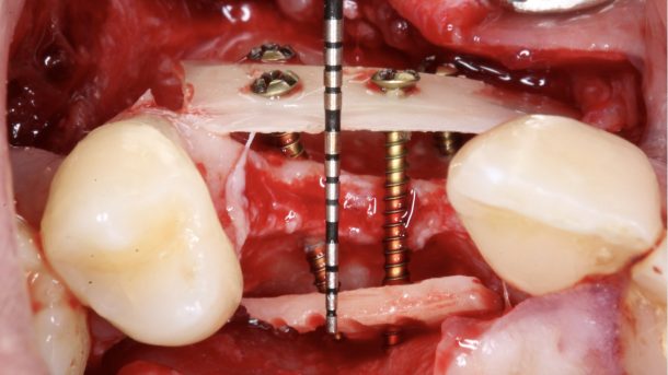
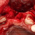
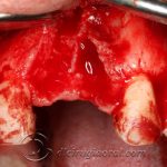
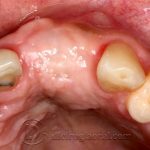
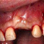
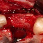
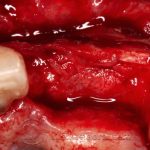
We made an intracrevicular incision with vertical incisions distant from the graft. Following a subperiosteal detachment we observed the good appearance of the graft which appeared well vascularised. We removed the fixing screws and drilled for the placement of implants.
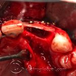
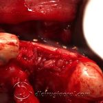
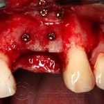
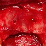
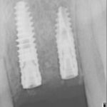
We could see the bone quality in the drill, which was difficult to drill, and inserted the implants in central and lateral incisor positions.
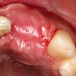
We sutured and waited another 3 months for osteointegration. In the future, we will perform the second stage with connective tissue graft and refer the patient to our prosthodontist colleague.
We hope you find the photographs interesting. The first 6 are of the previous reconstruction surgery.
-

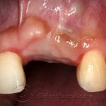
- Initial situation
-
- Defect occlusal view
-
- Defect
-
- Reconstruction occlusal view
-
- Filling in graft
-
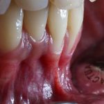
- Everything closed after 4 months
-
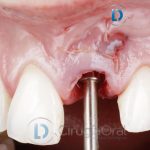
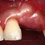
- Occlusal view
-
- Incision
-
- Appearance of graft
-
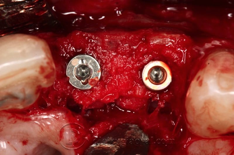
- Screws completely covered by bone
-
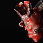
- Bleeding bone
-
- Occlusal view
-

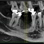
- Implant parallelism
-
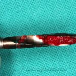
- Bone from drill
-
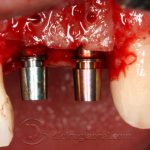
- Implants placed
-
- Intraoperative plate
-
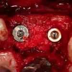
- Occlusal view
-
- Occlusal view
-
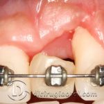
- Scarring
Click to rate this post!
[Total: 0 Average: 0]